


Management of Rhinophyma using 4 MHz Radiofrequency Technology | Application Report
Surgery remains a key treatment option for rhinophyma. An application report by Haneen Sadick, MD, of Mannheim University Hospital demonstrates the use of the CURIS® 4 MHz radiofrequency generator in combination with monopolar electrodes for tissue resection and nasal contouring. The report provides insights into the clinical procedure as well as the use of different electrodes for resection, coagulation, and contouring during an outpatient procedure.
- Published:
- Author: Sutter Medizintechnik GmbH
- Category: Precision in action | Our products in use
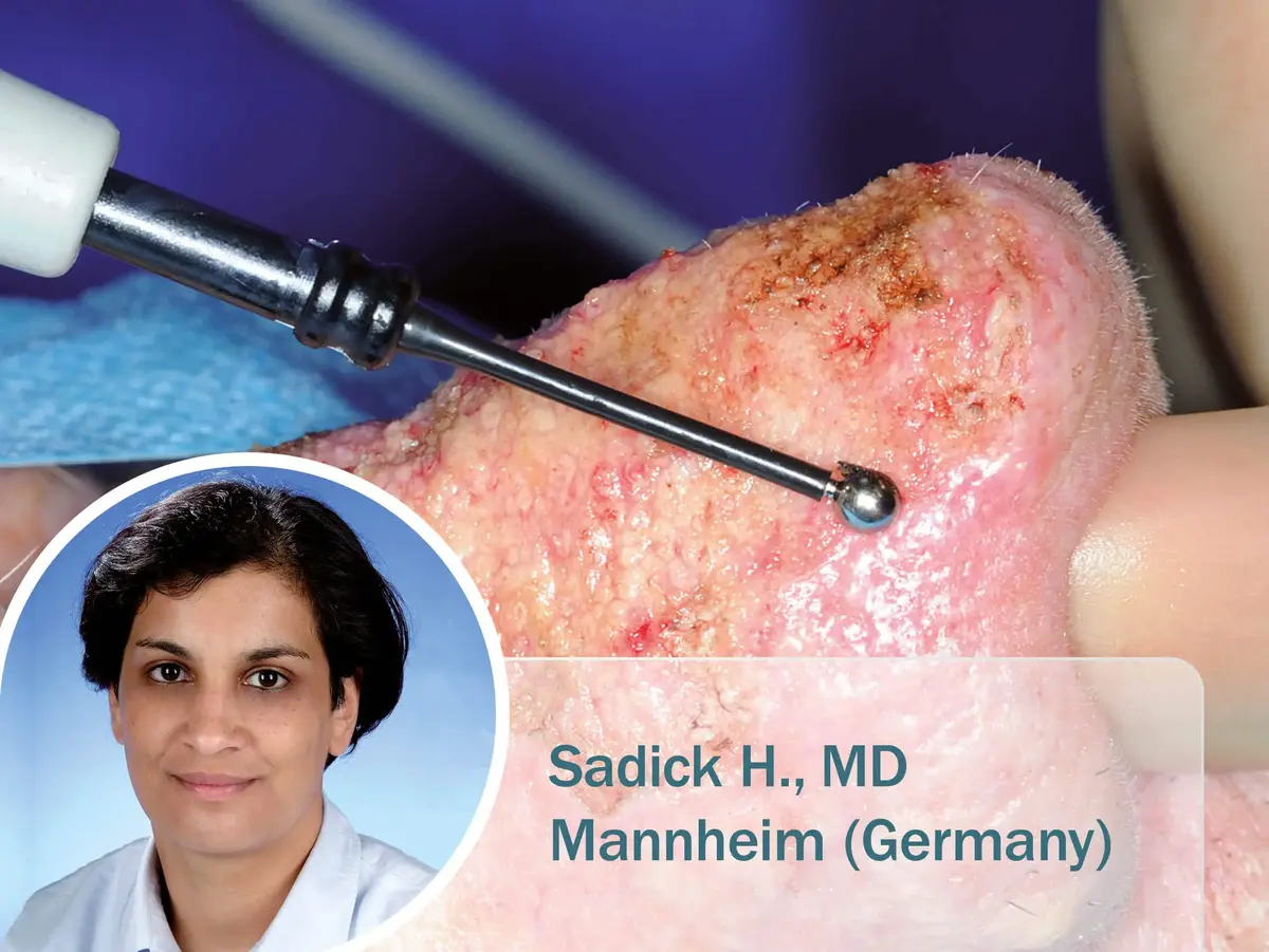
Rhinophyma is one of the most pronounced tissue changes affecting the nose and can be treated surgically. An application report by Haneen Sadick describes the use of radiofrequency surgery on the nose with the impedance-controlled CURIS® 4 MHz radiofrequency generator and monopolar electrodes in a patient with rhinophyma at the University Medical Center Mannheim, Germany.
Key Facts of the Application Report
- Topic: Management of rhinophyma using radiofrequency surgery of the nose
- Author: Haneen Sadick, MD
- Institution: Department of Otorhinolaryngology, Head and Neck Surgery, University Medical Center Mannheim, Mannheim, Germany
- Technology: CURIS® 4 MHz radiofrequency generator by Sutter
- Instruments: Monopolar wire loop electrodes and a monopolar ball electrode by Sutter
- Procedure Setting: Outpatient intervention performed under local anesthesia
- Follow-up: Regular outpatient follow-up examinations over a period of two months
What is Rhinophyma?
Rhinophyma is characterized by a slowly progressive enlargement of the lower third of the nose. Typical features include irregular thickening and nodular changes of the hypertrophic nasal skin. Although the underlying bony and cartilaginous nasal framework is not affected, the aesthetic contours of the nose can become significantly distorted. In addition, functional impairments, such as restricted nasal breathing, may occur. Surgical treatment presents particular challenges. Due to the rich vascular supply of the nose, intraoperative bleeding can occur, potentially compromising visibility within the surgical field.
Case Report from Mannheim: Radiofrequency Surgery for Rhinophyma
In the reported case, a 75-year-old patient presented to the Department of Otorhinolaryngology at the University Medical Center Mannheim with a progressive enlargement of the nose that had developed over several years. Clinical examination revealed marked hypertrophy of the sebaceous glands and subcutaneous tissue structures, particularly in the area of the nasal tip and the nasal alae.
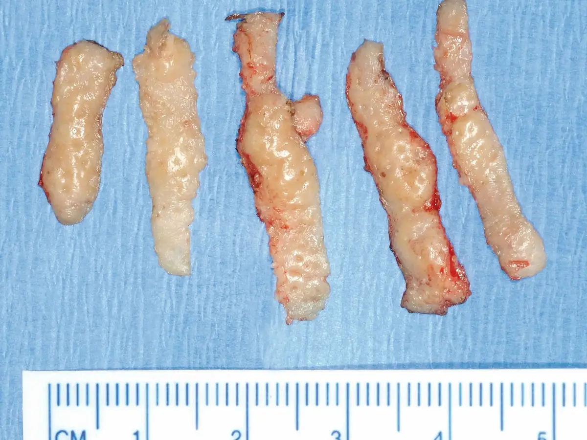
The treatment was performed on an outpatient basis under local anesthesia. A CURIS® 4 MHz radiofrequency generator was used in monopolar mode. Tissue resection was carried out step by step using a wire loop electrode to remove the excess tissue down to a clinically normal skin level. Particular care was taken to preserve the pilosebaceous units in order to minimize the risk of postoperative scarring.
Resection and Contouring with Monopolar Electrodes
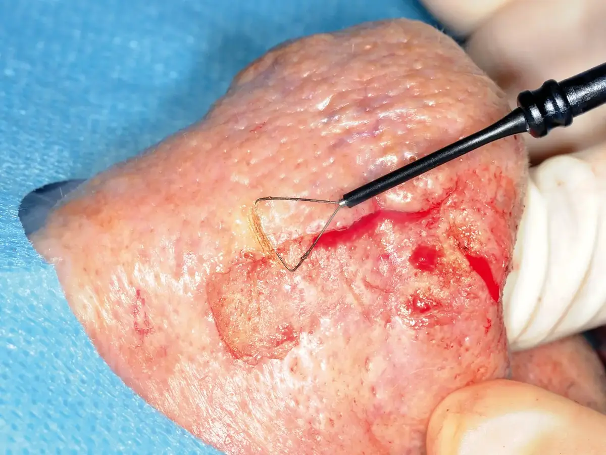
A triangular wire loop electrode and a round ball electrode were used for tissue resection. Following the removal of excess tissue, the nasal contour was refined using a 3 mm ball electrode to smooth surface irregularities and restore a natural appearance.
The application report highlights the ability to combine monopolar cutting and coagulation functions within a single procedure. This approach supports effective hemostasis while maintaining excellent visibility of the surgical field throughout the intervention.
“Radiofrequency surgery in the treatment of rhinophyma has proven to be an easy-to-handle, fast and efficient treatment modality. The combination of monopolar cutting and coagulation at the same time not only facilitates the re-shaping and sculpturing of the nose but also guarantees gentle hemostasis with excellent visibility of the surgical field.”
Haneen Sadick, MD, University Medical Center Mannheim, Germany
Follow-up Observations
According to the application report, the procedure was well tolerated by the patient. Regular outpatient follow-up examinations were conducted throughout the two-month observation period. Re-epithelialization of the nasal skin began approximately two weeks after the intervention. In this case, no wound infections, scar formation, or pigmentary changes were observed during follow-up.
As this is a single case report, these observations are limited to the patient described and should not be interpreted as representative of outcomes in all patients undergoing treatment for rhinophyma.
Instruments and Technologies Used
For the treatment of rhinophyma, a combination of a radiofrequency generator and specially designed monopolar instruments was used. The different components supported the individual treatment steps, from tissue resection to contouring of the nasal surface.


FAQ on the Management of Rhinophyma Using Radiofrequency Surgery
Rhinophyma is a progressive enlargement and thickening of the nasal skin that can occur as a severe manifestation of rosacea.
Surgical treatment is often considered when rhinophyma causes significant enlargement and thickening of the nasal skin, leading to distortion of the nasal contour and, in some cases, functional impairment such as obstructed nasal breathing. Surgery aims to remove excess tissue, restore the natural shape of the nose, and improve both aesthetic appearance and nasal function.
It is used for tissue resection and shaping of the nasal contour. At the same time, cutting and coagulation functions can be utilized simultaneously.
Monopolar wire loop electrodes were used for tissue resection, while a monopolar ball electrode was employed for contouring the nasal surface.
Particular care was taken to preserve these structures in order to minimize the risk of scar formation.
In the presented case, re-epithelialization was observed within two weeks, and no wound infections, scarring, or pigmentary changes were reported during the follow-up period.
The application report was authored by Haneen Sadick, MD, from the Department of Otorhinolaryngology, Head and Neck Surgery, University Medical Center Mannheim, Mannheim, Germany.